|
ULTRASOUND OF ECHOGENIC INTRACARDIAC FOCI |
- Small brightly echogenic foci without distal acoustic shadowing (1-3)
|
|

- Echogenicity is similar to or greater than surrounding bone (3). Should not be called an echogenic intracardiac focus if the echogenicity is less than bone and no defect is demonstrated on the color flow images.
|
|
|


- Do not produce acoustic shadowing.
|
|
|


- Occasionally referred to as a "golf ball".
- Exclude hyper-echogenicity of the ventricular walls or the moderator band in the right ventricle as the cause.
- An echogenic intracardiac focus has been associated with an increased risk of Down syndrome and trisomy 13 (8), trisomy 18 and monosomy X (9-12).
- Found in 2% of normal fetuses.
- Bromley and co-workers 1995(13).
- Incidence in normal fetuses = 4.8%
- Incidence in Down syndrome = 18%
- Increased risk of Down syndrome = 4-fold.
- Location (1).
- Left Ventricle (92.8%)
- Anterior papillary muscle.
- Cardiac apex.
- Interventricular septum.
- Posterior papillary muscle.
|
|
|

- Right Ventricle (4.8%)
- Anterior papillary muscle.
- Left and Right Ventricles (2.4%).
- Location within the atria have not been reported.
- Location has been suggested as an important factor in the risk of aneuploidy (7).
-
Normal karyotype (88% in
-
Aneuploid fetuses
(78% in
- Incidence of Down syndrome is higher among fetuses with RV or bilateral echogenic foci).
- Usually seen at the mid-trimester scan but can be seen at 12-13 weeks of gestation.
|
|
|
|
|
|
|
|
|

- Size.
- Usually 1-4 mm.
- Largest benign echogenic intracardiac focus reported is 6mm.
- 36% increase in size over time, 12% decrease and 51% remain unchanged (1).
- Complete resolution at the time of neonatal echocardiography in (40-48% of cases (2,4).

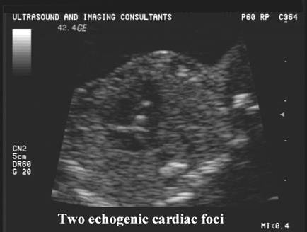
- Single or multiple (6-11%) (5,6).
|
|
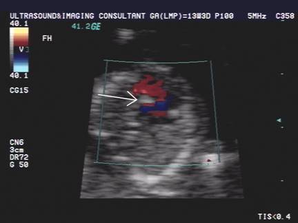
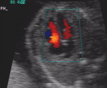
Single
EIF in left ventricle |
|
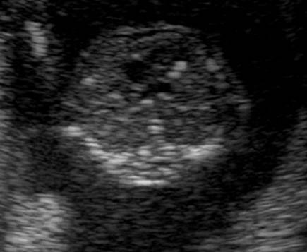
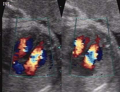
Two echogenic EIF left ventricle |
|
|
|
|
|
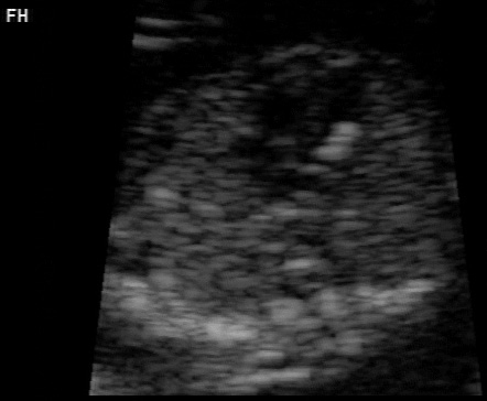
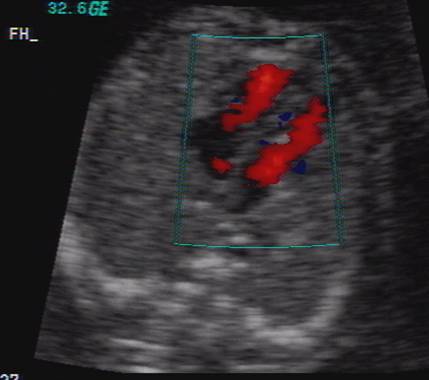
Multiple EIF left ventricle |
|
|
|
|


- Move synchronously with the valve leaflets throughout the cardiac cycle.
- Unusual echogenic foci:
- Diffuse echogenic
areas of the endocardium, interventricular septum, ventricular wall or
atrioventricular valves in 9/44 second trimester fetuses with echogenic
foci (8).
7 fetuses had adverse pregnancy outcome, 4 had cardiac dysfunction or heart defects, one case of missed abortion, metabolic disease and trisomy 13. - 9/65 second trimester fetuses (6) had echogenic foci of unusual size, shape, number, structure or location. Three had foci of 7-9 mm, four had multiple foci in the ventricles, one had a "double focus" and one had a "triple" focus. No association in this study of heart disease, chromosomal abnormality or poor outcome.
|
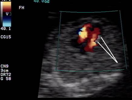
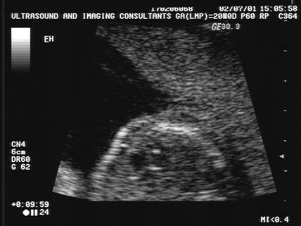
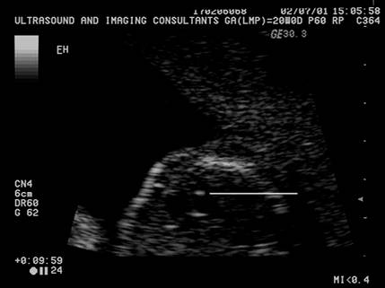
Echogenic
Intracardiac focus in the left ventricle. Note, by playing with the gain settings one can determine
that the density of the echogenic focus is similar to that of bone. |
|
|
|
|
|
|
|


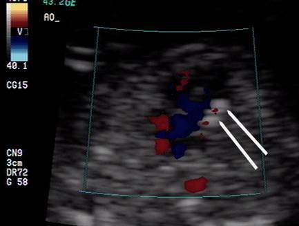
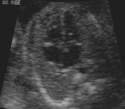
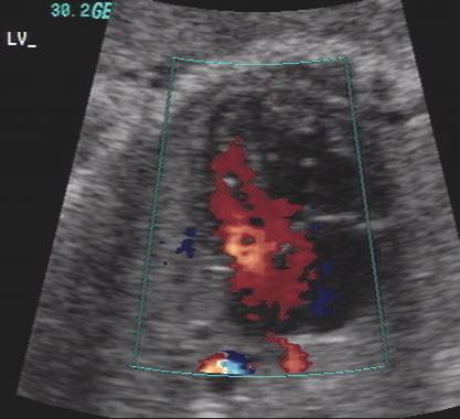
- Echogenic focus best visualized on the apical four chamber view. It may be hidden in the specular reflections of the ventricles in the subcostal four chamber view.
|
|
|


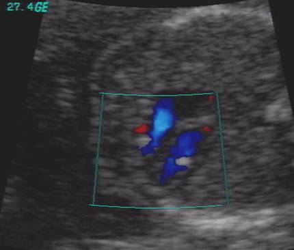
- An echogenic linear band may be seen in the ventricles and may cause a defect in the color flow, but should not be called an intracardiac focus.
These are not true echogenic intracardiac foci
|
|
|
|
|
|
|
|
Small
defects in the color flow
|
|
|
|
|
|
|
|

Roberts
and Genest (1992) (12). Prospective analysis of histological sections of 415
fetuses, found that a discrete central papillary muscle calcification was
significantly higher in fetuses with chromosomal aneuploidy (16% of fetuses
with trisomy 21 and 39% of fetuses with trisomy 13 versus 2% of control cases).
At this time it is not certain whether papillary muscle calcification is always
evident as an echogenic focus on ultrasound.
|
Ref |
MA (yrs) |
Ga (wks) |
Location |
Other ultrasound findings |
|
14 |
Not stated |
Not stated |
LV |
None |
REFERENCES |
- Petrikovsky BM, Challenger M, Wyse LJ. Natural history of echogenic foci within ventricles of the heart. Ultrasound Obstet Gynecol 1995;5:92-94.
- How HY, Villafane J, Parihus RR et.al. Small hyperechogenic foci of the fetal cardiac ventricle: a benign finding? Ultrasound Obstet Gynecol 1994;4:205-207.
- Romero D, Sepulveda W. Significance of echogenic foci in the fetal heart. Ultrasound Obstet Gynecol 1998;12:445-449.
- Simpson LM, Cook A, Sharland G. The significance of echogenic foci in the fetal heart: a prospective study of 228 case. Ultrasound Obstet Gynecol 1996;8:225-228.
- Petrikovsky B, Challenger M, Gross B. Unusual appearances of echogenic foci in the fetal heart: are they benign? Ultrasound Obstet Gynecol 1996;8:229-231.
- Bromley B, Lieberman E, Shipp T et.al. Echogenic Intracardiac focus (EIF): Association with aneuploidy in both high anf low risk patients. J Ultrasound Med;
- Brown DL, Roberts DJ, Miller WA. Left ventricular echogenic focus in the fetal heart, pathologic correlation J Ultrasound Med 1994; 13:613-616.
- Speulveda W, Cullen S, Nicolaidis P et.al. Echogenic foci in the fetal heart: a marker for chromosomal abnormality. Br J Obstet Gynaecol 1995;102:490-492.
- Twining P. Echogenic foci in the fetal heart: incidence and association with chromosomal disease. Ultrasound Obstet Gynecol 1993;3(Suppl. 2):175.
- Lehman CD, Nyberg DA,
Winter TC et.al. Trisomy 13 syndrome, prenatal
- Roberts DJ, Genest D. Cardiac histologic pathology characteristics of trisomies 13 and 21. Hum Pathol 1992; 23:1130-1140
- Bromley B, Lieberman E, Laboda LA, Bernacerraf BR. Echogenic intracardiac focus, a sonographic sign for Down syndrome. Obstet Gynecol 1995; 86:998-1001
- Schechter AG, Fakhry J, Shapiro LR et.al. In utero thickening of the chordae tendinae. A cause of intracardiac echogenic foci. J Ultrasound Med 1987;6:691-695.
- Simpson JM, Cook A, Sharland G. The significance of echogenic foci in the fetal heart: a prospective study of 228 cases. Ultrasound Obstet Gynecol 1996;8:225-228.
- Bromley B, Lieberman E, Shipp TD et.al. Significance of echogenic intracardiac intracardiac focus in fetuses at high and low risk for aneuploidy. J Ultrasound Med 1998;17:127-131.
- Manning JE, Ragavendra N, Sayre J et.al. Significance of fetal intracardiac echogenic foci in relation to trisomy 21: a prospective sonographic study of high-risk pregnant women. Am J Roentgenol 1998;170:1083-1084.