|
THE |
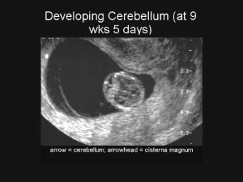
- Can be sonographically visualized as early as 9-10 weeks.

- It grows rapidly in the second trimester having a linear relationship with gestational age. Measurement in mm equals approximate gestational age in weeks.
- Peanut shaped with a central constriction denoting the vermis and flared ends representing the two hemispheres.
- Its location in the posterior fossa (surrounded by the dense petrous ridges and occipital bone) makes it more resistant to deformation by extrinsic pressure.
- It has therefore been proposed that the Transcerebellar diameter (Table / Graph) is a better predictor of gestational age than the BPD when there are variations in the shape of the fetal head (dolicocephaly or brachycephaly).
|
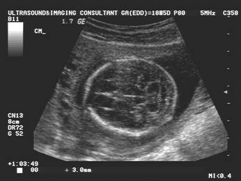
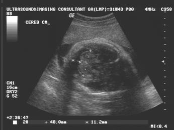
Transcerebellar Diameter |
Cisterna magna |
|
|
|

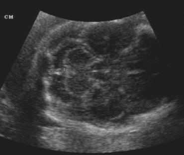
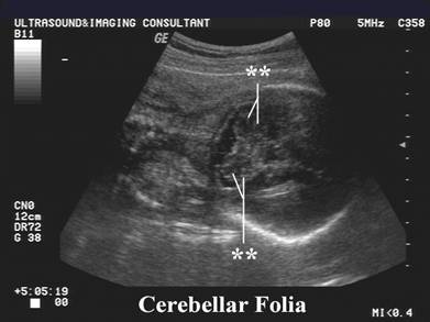
· Sonographic changes in the appearance of the cerebellum throughout gestation.
Sonographic appearance of the cerebellum through gestation |
|
|
|
Grade 1:
|
|
|
Grade II:
|
|
|
Grade III:
|


NORMAL VARIANT – DEFECT IN THE INFERIOR
VERMIS
|
Normal Variant - Defect in the Inferior Vermis |
|||
|
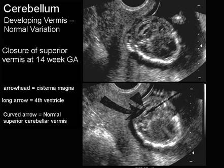
During normal embryologic development of the
posterior fossa the cerebellar vermis appears to form by fusion of the
cerebellar hemispheres superiorly and in the midline during week 9 of
gestation (1,2). Fusion continues inferiorly with complete
closure of the vermis being complete at the end of the 15th week. Vermian development is one of the last steps in the
formation of the cerebellum occurring when crown-rump length is 150 mm (2).
|
In a recent study (3), 56% of fetuses had an open vermis
at 14 weeks gestation, decreasing to 23% at 15 weeks and 6% at 17 weeks.
Therefore a diagnosis of Dandy-Walker variant should not be made prior to 18
weeks gestational age. |
||
|
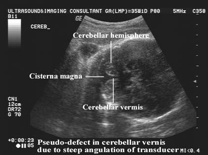
Normal Variant – Defect in Inferior Vermis created by a steep
transducer angle. |
|
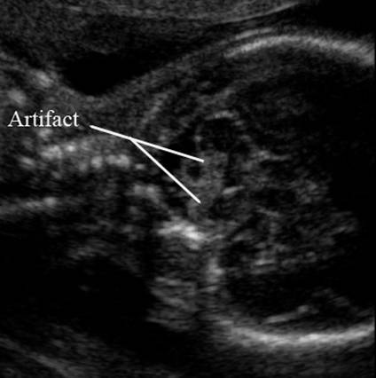
||
|
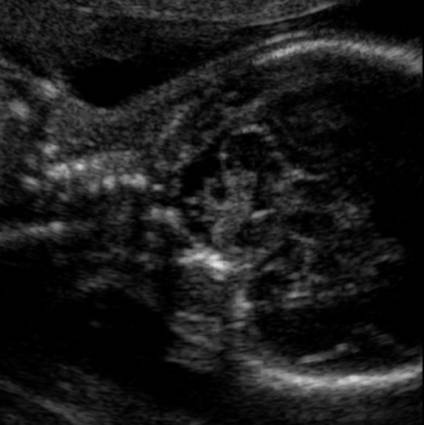
Echogenic band
of tissue crossing the vermis - artifact |
|
||


REFERENCES
|
- Barkovich AJ, Kjos BO, Norman D et.al. Revised classification of posterior fossa cysts and cystlike malformations based on the results of multiplanar MR imaging. AJR 1989;10:977-988.
- Lemire
RJ, Loeser JD, Leech RW et.al.
Normal and abnormal development of the human nervous system.
- Bromley B,