|
ABSENT FETAL URINARY
BLADDER BLADDER AGENESIS |
The bladder may be seen as early as 10 weeks of gestation but it is not reliably visualized until 13 weeks.
If the bladder is not visualized during a 30 min. examination a repeat scan at 60 and 90 minutes should be obtained.
FREQUENCY OF VISUALIZATION |
Frequency of Visualization of the Bladder VS Gestational Age
TRANSIENT NON-VISULAIZATION |
Non- visualization is usually a transient finding, especially in the presence of normal amniotic fluid volume. These patients should be rescanned in a few day to confirm a normal bladder is present. It is always important to exclude bladder exstrophy when the bladder is persistently not visualized in the presence of normal non obstructed kidneys and normal amniotic fluid volume.
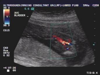
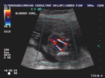
Absent bladder at 18 wks 5 days during the entire
scan. amniotic fluid
volume. Normal bladder between the
umbilical arteries at 20 wks 5 days.
PERSISTENT NON-VISUALIZATION |
- Bladder agenesis –
The bladder
develops from the vesical portion of the urogenital sinus and the trigone
from the inferior ends of the mesonephric ducts.
Bladder absence is rare and incompatible with life. If seen, it is often
part of a complex anomaly in a stillborn fetus. Cloacal
development is possibly disturbed by lack of a bladder distended by urine
and results in failure of incorporation of ducts and ureters
into the trigone. Fetal bladder absence may be
simulated by recent emptying requiring follow-up imaging for confirmation
of bladder absence. An axial
- Bilateral
renal agenesis.
- Bilateral
multicystic renal dysplasia.
- Unilateral
renal agenesis + contralateral multicystic renal dysplasia.
- Unilateral
renal agenesis or multicystic renal dysplasia + contralateral
severe renal obstruction or dysplasia.
- Severe
bilateral renal obstruction or dysplasia.
- Severe
autosomal recessive polycystic disease of the
kidney.
- Severe
IUGR.
- Persistent cloaca.
- Cloacal exstrophy.
- Bladder exstrophy.
REFERENCES
|
- K.L.
Moore and T.V.N. Persaud. The Developing Human.
Clinically Oriented Embryology (ed 7.), Saunders,
- T. Berrocal, P. Lopez-Pereira, A. Arjonilla et al., Anomalies of the distal ureter, bladder and urethra in children: Embryologic, radiologic and pathologic features. Radiographics 22 (2002), pp. 1139–1164.