|
ULTRASOUND OF
HYDROCOLPOS / HYDROMETROCOLPOS |
- Female fetus.
- The pathognomonic physical finding in persistent cloaca is a single perineal orifice between the labia minora is probably not possible to demonstrate sonographically.
- Usually a hypoechoic or anechoic pelvic mass which may contain low level echoes (1,2).
|
|
- Occasionally a homogeneous midline pelvic mass (3).
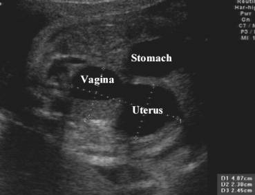
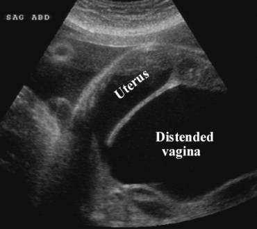
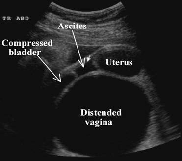
- Distended vagina situated posterior to the bladder and anterior to the rectum. The uterus is rarely involved due to the ability of the vagina to distend (4).
- Membrane bulging through the perineum and spreading the labia majora (2).
- ± Cervical distention.
- ± Uterine distention.
- ± Fluid-debris level (distended vagina).
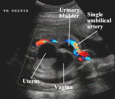
- Bladder often not identified (compression by distended vagina).
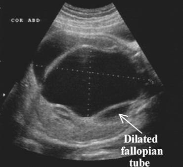
- Duplication of the uterus and vagina in 41%.
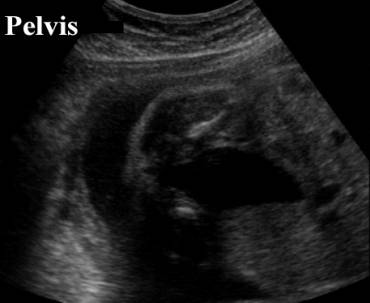
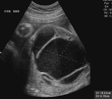
Cystic mass in pelvis extending up to perineum (22 wks GA) |
|
|
|
|
|
|
|
|
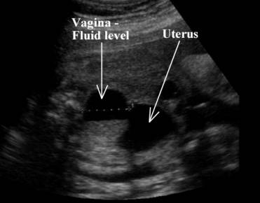
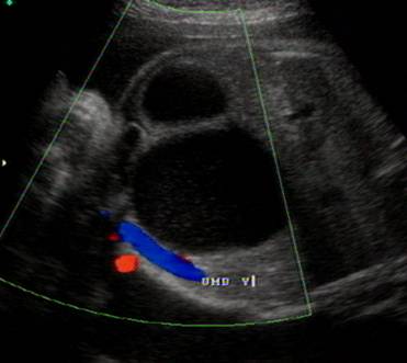
Fluid level in
vagina Communication between vagina and uterus |
|
|
|
|
|
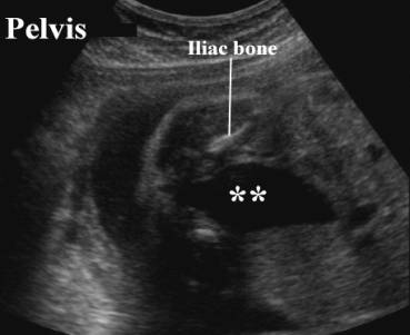
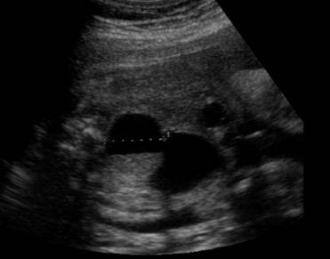
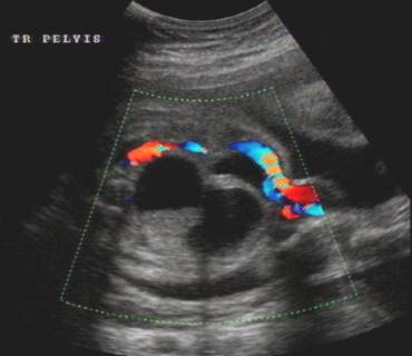
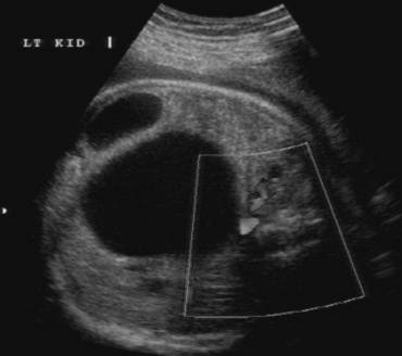
Bladder
compressed by distended vagina Single umbilical artery |
|
|
|
|
|
|
|
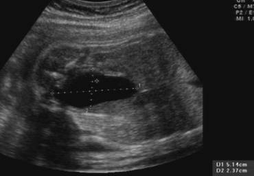
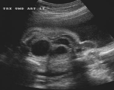
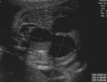
Uterus and vagina |
|
|
|
|
|
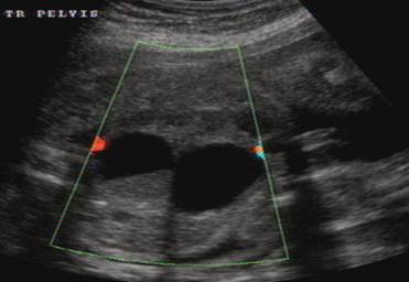
Scan at 32 weeks of gestation |
|
|
|
|
|
|
|
|
Acsites |
|
|
|
|
REFERENCES |
- Davis GH, Wapner
RJ,
- Winderl LM, Silverman RK. Prenatal diagnosis of congenital imperforate hymen. Obst Gynecol 1995;85(5):857-860.
- Hill SJ, Hirsch JH. Sonographic detection of fetal hydrometrocolpos. J Ultrasound Med 1986;5:211-213.
- Kay R, Tank ES. Principles of management of persistent cloaca in the female newborn. J Urol 1977;117:102-104.