|
RETROPERITONEAL
LYMPHANGIOMA |
Lymphangiomas are benign hamartomas
of the lymphatic system, consisting of multiple dilated lymphatic channels.
Although they usually occur in infancy, childhood or adolescence (1), they have
been reported antenatally (2). Lymphangiectasia
results due to the pressure of lymph accumulating in anomalous dilated
lymphatic vessels. They are believed to arise from a developmental defect in
the lymphatic pathways, which usually develop from the 6th week of
gestation. Failure of the embryonic lymph sacs to establish communication with
the venous system or aberrant budding of the primordial lymph sacs has also
been suggested as the etiology.
CLASSIFICATION |
- Simple (simplex) – capillary size channels.
- Cavernous – dilated lymphatic channels frequently with a fibrous adventitial covering.
- Cystic (uni or multilocular containing serous, chylous or bloody contents) – lined by endothelial cells.
- Mixed lesions may co-exist in different areas of the same lymphangioma.
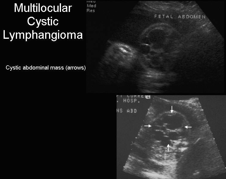
ULTRASOUND (1-11) |
- Cystic abdominal mass.
- Simple or multiseptate.
- Septa are thin (1-2mm).
- A rim of calcification may be present but this is unusual (1).
- Fine echoes within the cyst may be due to hemorrhage.
- Left sided tumors are more common.
|
|
- To date there are numerous reported cases in the literature. A significant number showed extension down the left leg. This extension is thought to reflect the pathway of the normally developing lymphatic system and may therefore be typical of retroperitoneal lymphangiomas.
- Hydronephrosis due to extrinsic ureteric obstruction (2).
- Bowel obstruction is very rare (3).
- Hydrops and polyhydramnios may be present.
|
Isolated antenatal abdominal lymphangiomas |
|||
|
Ref |
GA at diagnosis |
Ultrasound
findings |
Outcome |
|
4 |
19 wks |
Left sided multilobulated mass extending into leg |
Pregnancy termination |
|
6 |
24 wks |
Left sided, multiseptated, anechoic mass |
Complete surgical resection at 2 months |
|
7 |
28 wks |
Multiple cystic masses, left abdomen |
Stable; surgical resection at 1 yr if unresolved |
|
8 |
28 wks |
Right sided multiseptated mass; extension into thigh |
Pregnancy termination |
|
9 |
31 wks |
Right sided, very large multiseptated cystic mass |
Excised completely at 6 days of age |
|
10 |
31 wks |
Massive cystic mass extending from left chest wall to buttock |
Postnatal sclerotherapy. Partly successful |
|
5 |
20 wks |
Left sided multicystic septated mass extending to buttock and thigh |
Pregnancy termination |
|
11 |
28 wks |
Left multilobulated cystic mass |
Complete excision at 3 months of age |
|
12 |
19 wks |
Multiloculated cystic mass on left side |
Excision at 6 wks of age due to abdominal distention and vomiting. |
DIFFERENTIAL DIAGNOSIS |
- Cystic teratoma (ovarian or retroperitoneal).
- Enteric duplication cyst.
- Mesenteric cyst.
- Meconium pseudocyst.
- Choledochal cyst.
- Cystic renal tumor.
- Urachal cyst.
REFERENCES |
- Davidson AJ, Hartman DS. Lymphangioma of the retroperitoneum: CT and sonographic characteristics. Radiology 1990;175:507.
- Malnofski MJ, Poulton TB, Nazintsky K et.al. Prenatal ultrasonic diagnosis of retroperitoneal cystic lymphangioma. J Ultrasound Med 1993;12:427-429.
- Blumhagen JP, Wood BJ, Rosenbaum DM. Sonographic evaluation of abdominal lymphangiomas in children. J Ultrasound Med 1987;6:487.
- Kozlowski HJ, Frazier CN, Quirk JG Jr. Prenatal diagnosis of abdominal cystic hygroma. Prenat Diagn 1988;8(6):405-409.
- Deshpande P, Twining P, O’Neill D. Prenatal diagnosis of fetal abdominal lymphangioma by ultrasonography. Ultrasound Obstet Gynecol 2001;17:445-448.
- Devesa R, Munoz A, Torrents M et.al. Prenatal ultrasonographic findings of intra-abdominal cystic lymphangioma: a case report. J clin Ultrasound 1997;25(6):330-332.
- Katz VL, Watson WJ, Thorp JM Jr et.al. Prenatal sonographic findings of massive lower extremity lymphangioma. Am J Perinatol 1992;9(2):127-129.
- Kaminopetros P, Jauniaux E, Kane P et.al. Prenatal diagnosis of extensive fetal lymphangioma using Sonography, magnetic resonance imaging and cytology. Br J Radiol 1997;70(835):750-753.
- Suzuki N, Tsuchida Y, Takahashi A et.al. Prenatally diagnosed cystic lymphangioma in infants. J Pediatr Surg 1998;33(11):1599-1604.
- Suchet I. Unpublished case.