|
SONOGRAPHIC
ASSESSMENT OF CARDIAC FUNCTION |
DOPPLER ASSESSMENT
|
|
1.
Principles 4.
Doppler findings in growth retarded
fetuses 5.
Doppler findings in congenital heart
disease 6.
Doppler findings in fetal anemia 7.
Doppler findings in fetal non-immune
hydrops |
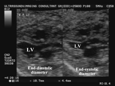
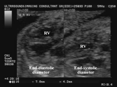
Systolic Function
LV ejection fraction:
Shortening
fraction: LVED – LVES / LVED X100 = 34%
+/- 3%
end-diastolic dimension – end-systolic dimension divided
|
|
|
|
Ventricular Diastole LVED = Left ventricular end-diastole |
|
|
Ventricular Systole LVES – Left ventricular end-systole |
|
|
|
|
Diastolic
Function. ·
Venous flow patterns: o IVC / hepatic
veins. o Ductus
venosus. o Umbilical
vein. ·
Inflow (mitral and tricuspid valves). |
|
|
|
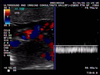
Normal waveform in the umbilical vein
|
|
|
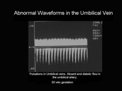
Abnormal waveform in the umbilical vein - notching |
|
|
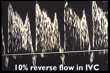
Normal waveform in the inferior vena cava |
|
|
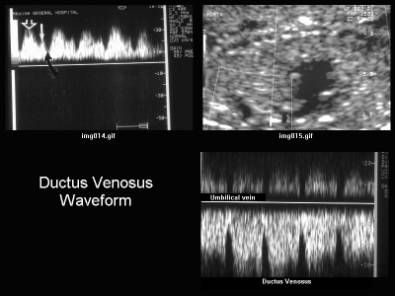
Normal waveform in the ductus venosus |
|
|
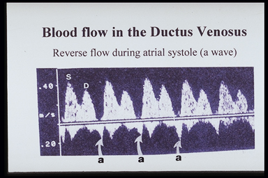
Abnormal waveform in the ductus venosus – reversal of the A
wave. |