|
ULTRASOUND OF ATRIAL
SEPTAL DEFECTS |
This is a difficult antenatal diagnosis because of the normal physiological presence of the foramen ovale.
An anechoic area is present in the atrial septum best seen in the four-chamber view. A large left to right shunt is physiologic in utero, therefore the ASD does not compromise the fetus hemodynamically in utero.
- Septum primum has a configuration that resembles a "loose pocket", and may therefore appear as circular or linear depending on the plane of the section.
- Septum secundum is thicker and less mobile and makes up the bulk of the interatrial septum.
|
|
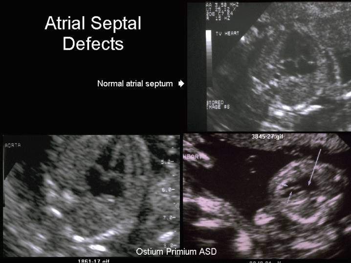
Normal Atrial
Septum
|
Ostium Primim Atrial Septal Defcet |
|
|
|
|
|
Ostium Secundum Atrial Septal Defect |
|
|
|
|


- Best visualized on subcostal four-chamber view of the heart.
- Ostium secundum ASD's appear as a larger than expected area of dropout in the central portion of the septum secundum (in the vicinity of the foramen ovale) or as a deficient foraminal flap (septum primum) that fails to cover the entire foramen ovale. There will still be visible tissue between the defect and the mitral/tricuspid valve leaflets and the back wall of the atria.
- Ostium primum ASD's result
in the absence of the lower portion of the atrial septum (just above the atrioventricular
valves). The septal leaflet of the tricuspid valve and anterior leaflet of
the mitral valve insert at the same level (normally the tricuspid valve
inserts slightly more apical than the anterior leaflet of the mitral
valve).
The normal "fish mouth" appearance of the mitral valve in the short-axis view is absent as the anterior leaflet of the mitral valve contains a large cleft. - Sinus venosus ASD's have not been reported antenatally.
|
|
Video clip of an Ostium
Primum Atrial Septal Defect
|
|
|
|
|
|