|
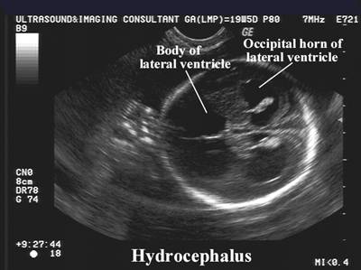
ULTRASOUND CRITERIA
FOR THE DIAGNOSIS OF
HYDROCEPHALUS |
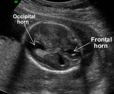
- Dilatation of the Posterior
Horn of the Lateral Ventricle
The posterior horn is the first portion of the lateral ventricle to dilate. - Normal values for the posterior horn
- The anterior horns of the lateral ventricles are the last to dilate.
|
|
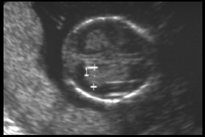
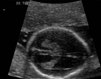
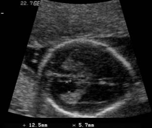
Dilatation of the Posterior Horn of the Lateral Ventricle |
|
|
|
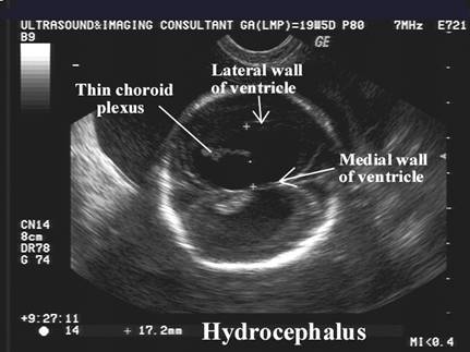
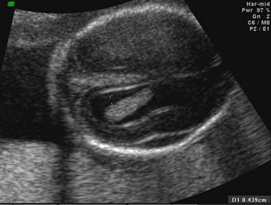
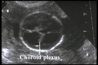
- Compression and thinning of the choroid plexus due to the increased CSF pressure.
|
|
Severe Hydrocephalus Note:
|
|
|
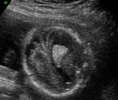
Normal Choroid Plexus at 18 wks GA. |
|
|
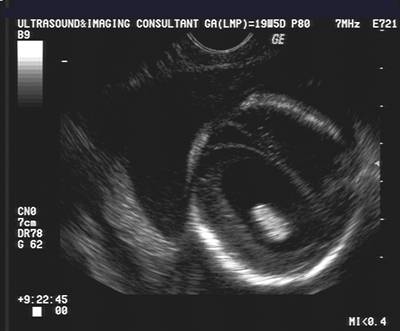
Thin Choroid Plexus and
Hydrocephalus at 18 wk GA. |
|
|
“Hanging”
choroids plexus sign in hydrocephalus |
|
|

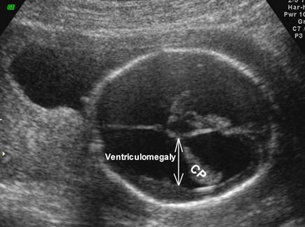
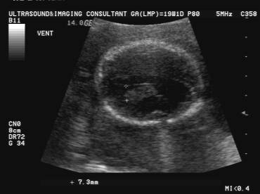
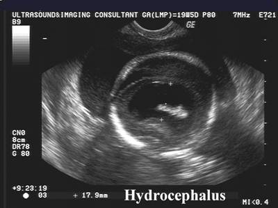
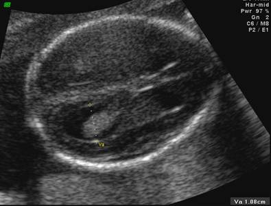
- Size of the lateral
ventricle at the level of the atrium.
Normal size of the ventricular atrium - <1 cm = normal.
- 1-1.2 cm = borderline ventriculomegaly
- >1.2-1.5 cm = mild ventriculomegaly
- >1.5 cm = marked ventriculomegaly
4. Borderline
ventriculomegaly.
|
|
|
Normal
ventricular atrium – 7 mm. Echogenic choroid plexus completely fills the ventricle at the the level of the atrium. |
|
Normal
Ventricles |
Borderline ventriculomegaly |
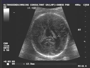
Marked
Ventriculomegaly (17.9 mm) |
|
|
|
|
|
|
|
|
|
|
|
|
|
|
|
|

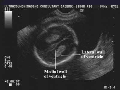
5. Hydrocephalus:
a. Ventricular atrium ³1.2 cm.
b. Medial separation of the choroid plexus from the medial wall of the lateral ventricle.
i. <3 mm = normal
ii. 3-5 mm = "gray zone".
iii. >5 mm = abnormal separation.
c. Hanging or dangling choroid plexus sign (the dependent choroid plexus hangs down towards the dependent part of the head by gravity.)
|
|
|
|
|
Hanging choroids plexus sign
|
6.
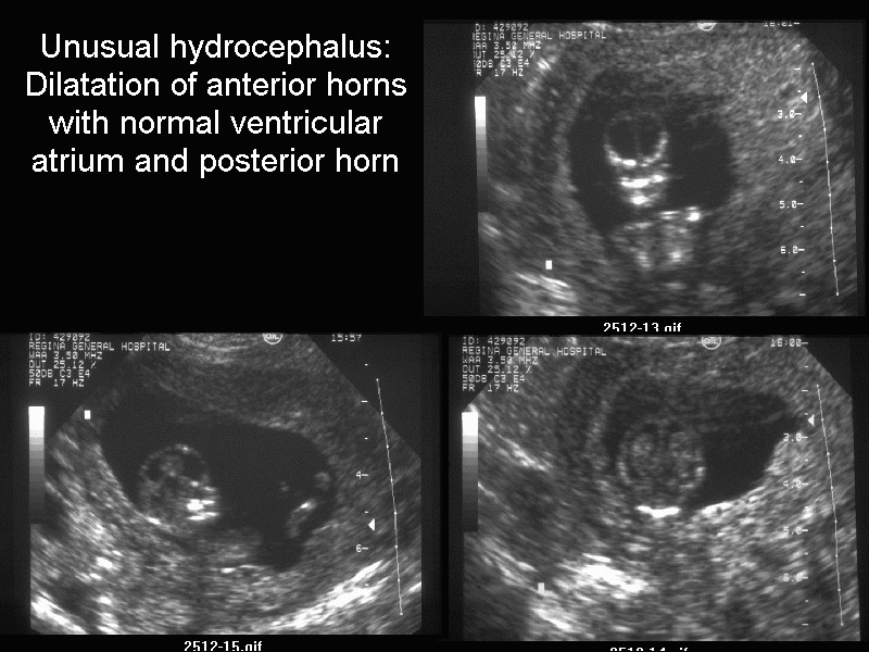
Unilateral /
Asymmetrical Hydrocephalus.
Detection or unilateral or asymmetrical hydrocephalus is difficult to diagnose unless the fetus is scanned through the sutures or fontanelles so that the near field ventricle is not obscured by reverberation artifact.
· Ventricular atrium > 10 mm.
· > 2 mm discrepancy between the two sides.
· Unilateral or bilateral dilatation with asymmetry in ventricular size ( > 2 mm).
· IF isolated, mild and stable in utero, favorable neurological outcome is common.
· If rapidly evolving or associated with other abnormalities, a poor neurological outcome can be expected.

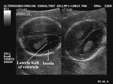
7. False positive diagnosis of ventriculomegaly – incorrect measurement of the echogenic line of the insula as the lateral wall of the ventricle.
|
Incorrect Measurement |
Correct Measurement |
|
|
|