|
NORMAL FACE |
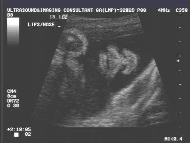
The face should be routinely evaluated in all second and third trimester fetuses. Optimal evaluation may be limited by fetal position however one should endeavor to obtain midsagittal profile and coronal sections of the lower facial bones.
· The forehead, orbits, nose, lips and ears can be consistently identified from 12 weeks of gestation.
· Sagittal, transverse and coronal planes are all useful for the evaluation of normal and abnormal anatomy.
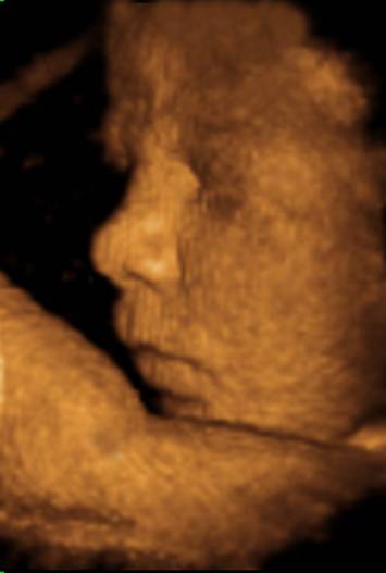
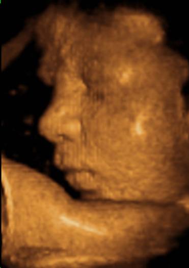
· A mid-sagittal plane allows visualization of the fetal profile.
|
|
|
|
|
|
|
|
|
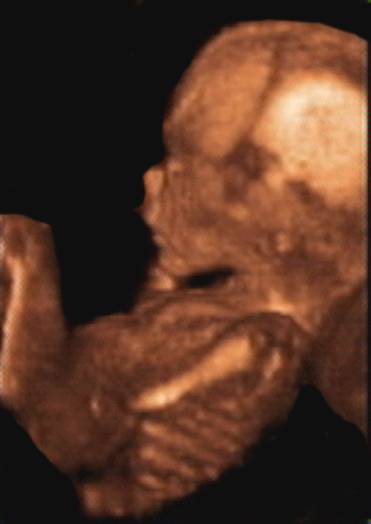
· The ears are visualized in parasagittal scans tangential to the calvarium.
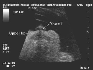
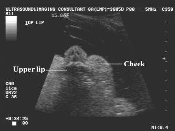
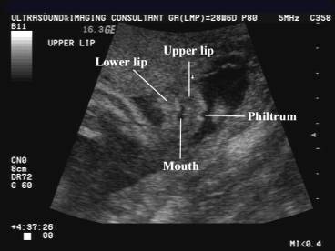
· The coronal planes are probably the most important ones in the evaluation of the integrity of facial anatomy.
· Orbits, eyelids, nose, and lips are well visualized.
· The tip of the nose, the alae nasi, and the columna are seen above the upper lip.
· The nostrils typically appear as two little anechoic areas.
·
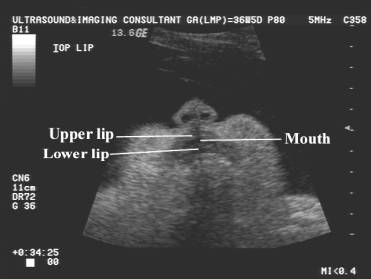
The anterior mandible is at the level of the lower lip,
which should be directly under the upper lip. This relationship is usually made
subjectively, however tables for Normal
Mandibular Length have been reported.
|
|
|
|
|
|
|
|
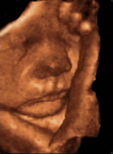
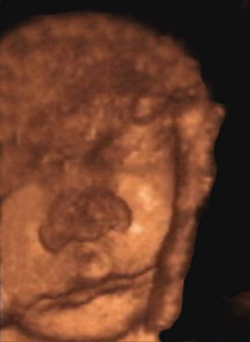
Note: a prominent philtrum may simulate a small cleft. 3D views help in differentiating a cleft from a normal philtrum. |
|
|
|