|
ULTRASOUND OF FIBULA
HEMIMELIA (1-6) |
- Unilateral (one thirds of cases).
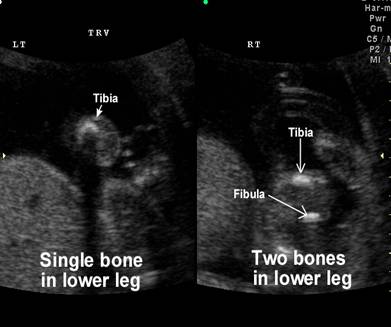
- Complete (more commonly) or incomplete absence of the fibula
- Unilateral in one-third of cases.
- Males more common than females.
- Degree of deficiency varies.
- Anteromedial bowing of the tibia (with/without shortening). There is a skin dimple at the point of greatest angulation.
- Talipes equinovalgus.
- Absence of one or more lateral rays of the foot (2).
- In bilateral involvement the tibia are usually straight.
- Non-skeletal anomalies are
rarely reported (0.8%)
- Ipsilateral shortening of the femur.
- Abnormal femoral head and neck (1).
|
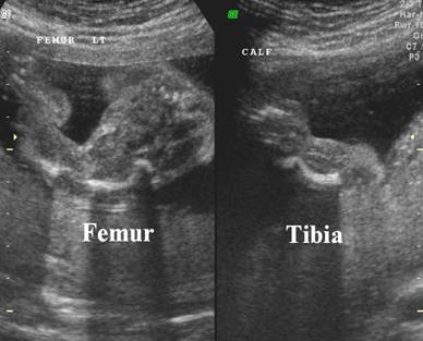
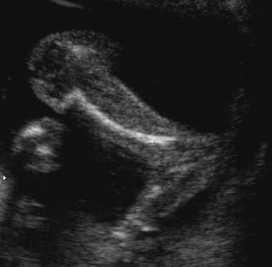
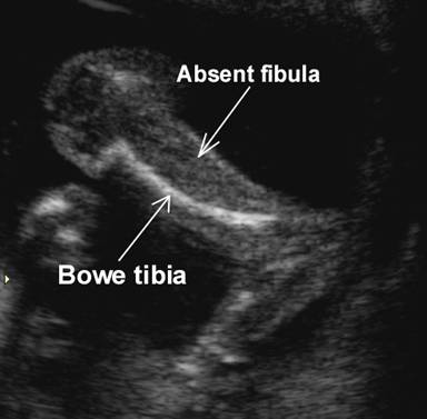
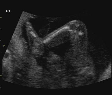
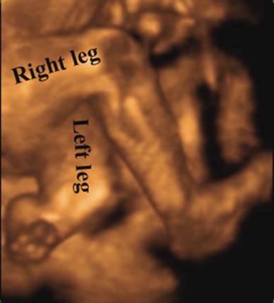
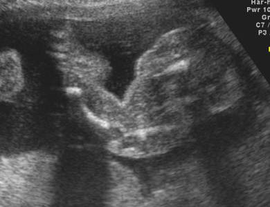
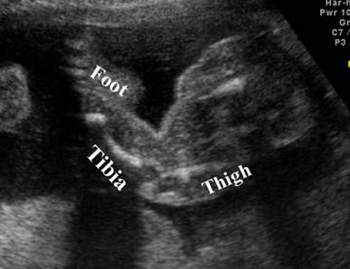
Case
1 - Bowing and shortening of the tibia and femur Absent fibula |
|
|
|
|
|
|
|
|
|
|
|
|
|
|
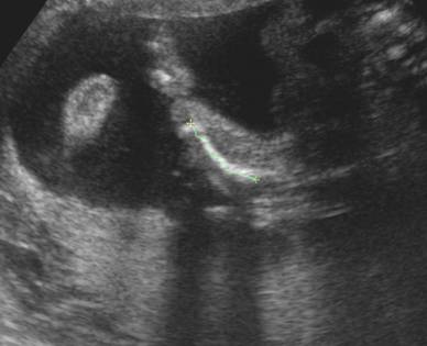
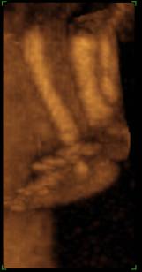
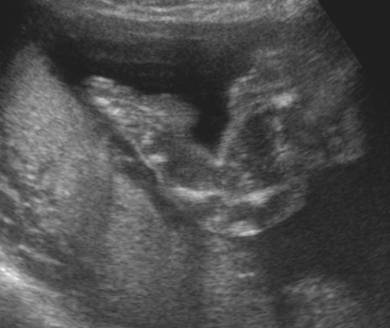
Case
2 – Absent fibula associated with proximal focal femoral deficiency |
|
|
|
|
|
|
|
|
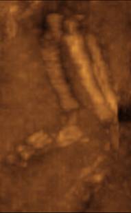
Talipes
equinovarus |
|
|
|
|


ASSOCIATIONS |
- Frequently associated with abnormalities of other bones, pelvis and extremities.
- Almost 50% of patients with
focal femoral deficiency have fibula hemimelia.
- Femur-fibula-ulna
syndrome (4).
- Omphalocele (5,6).
REFERENCES |
- Lewin SO, Opitz JM. Fibula hypoplasia: Review and documentation of the fibular developmental field. Am J Med Genet (Suppl) 1986;2:215.
- Froster
UG, Baird PA. Congenital defects of lower limb and associated
malformations: A population based study. Am J Med Genet 1993;45:60.
- Hirose
K, Koyanagi T, Hara K et.al.
Antenatal ultrasound diagnosis of the femur-fibula-ulna syndrome. J Clin Ultrasound 1988;16:199.
- Sepulveda
W, Weiner E, Bridger JE et.al. Prenatal
diagnosis of congenital absence of the fibula. J Ultrasound Med 1994;13:655-657.
- Uffelman J, Woo R, Richards DS. Prenatal diagnosis of bilateral fibular hemimelia. J Ultrasound Med 2000;19:341-344.