|
SACRAL LENGTH |
The sacral vertebrae ossify is a predictable manner (1), having three
primary ossification centers, one for the body (centrum), and one for each
posterior neural arch. The sacral can be easily and constantly visualized and
has therefore been reported to provide a reproducible landmark for measurement
(2).
ULTRASOUND |
|
|
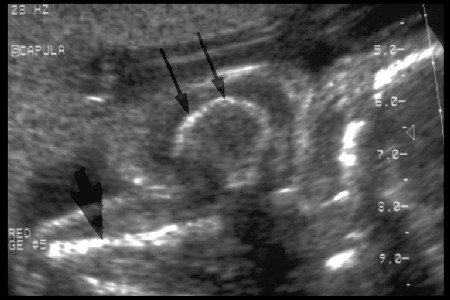
- Best visualized with the occiput anterior (in a vertex presentation), or the sacrum anterior in a breech presentation.
- Measurements are difficult when the spine is posterior and adjacent to the uterine wall.
- The coccyx is cartilaginous at birth making it easy to differentiate from the lowermost sacral segment (S5).
- Five sacral ossification centers in the centra can be visualized at 15 weeks gestation. Neural arch ossification is not seen before 22 weeks gestation (2).
- Measurement:
- Anterior aspect of the vertebral body (centrum)
- Measured in a sagittal plane from the anterosuperior aspect of S1 to the distal tip of the spine of S5.
- Do not include the unossified coccyx in the measurements (this may appear as a thin echogenic plate in the third trimester).
- Normal Sacral Length (Table).
- "Fractional spine length" (3) - distance of the longitudinal axis between T10 and L5. This measurement has been found to correlate strongly with femur length.
REFERENCES |
- Budorick NE, Pretorius DH, Grafe MR et.al. Ossification of the fetal spine. Radiology 1991;181:561-565.
- Sherer DM, Abramowicz JS, Plessinger MA et.al. Fetal sacral length in the ultrasonographic assessment of gestational age. Am J Obstet Gynecol 1993;168:626-633.
- Li DF, Woo JSK. Fractional spine length: a new parameter for assessing fetal growth. J Ultrasound Med 1985;5:379-383.