|
FETAL PELVIS IN DOWN SYNDROME |
The abnormal development of the pelvis in fetuses with Down syndrome is thought to be a primary developmental abnormality, although some workers have speculated on the influence or irregular growth and generalized hypotonic musculature (1).
Studies have shown that the fetal pelvis may be useful in detection of
trisomy 21 in the 15-20 week gestational age (2).
ULTRASOUND |
- X-Ray appearance of the pelvis has a typical appearance.
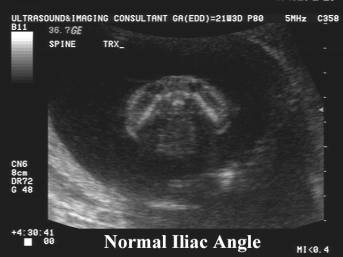
- Wing of ilium tends to flare laterally producing a wide iliac angle.
- Normal mean iliac angle = 60°.
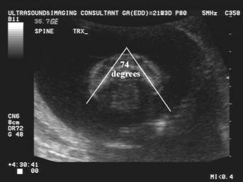
- Down syndrome mean iliac angle = 75°.
- Curvature of the wing of the ilium. Workers (2) were unable to detect a statistically significant difference between normal and fetuses with Down syndrome.
- Iliac wing angle (3). This is the angle between the left and right iliac wings measured on an axial transverse view of the fetal pelvis. It is measured at the level where the iliac bones are at their longest dimensions and are approximately equal. The angle is very dependent on the level at which it is measured. A larger angle is present if the measurement is more cephalad and decreases at a more caudal level.
- Fetuses with Down syndrome have a greater iliac wing angle than do normal fetuses.
- Using a cutoff value of 90°, the authors detected 90.9% 0f fetuses with Down syndrome with a specificity of 94.5% and a positive predictive value of 33% in their high-risk population.
- Iliac length. An observed-to-expected iliac length of ³1.21 for gestational age has a sensitivity of 40%, a positive predictive value of 2.6% and a false positive rate of 2% for the detection of Down syndrome (4).
REFERENCES |
- Kaufmann HJ, Taillard WF. Pelvic abnormalities in mongols. Br Med J 1961;5230:948-949.
- Kliewer MA, Hertzberg BS, Freed KS et.al. Dysmorphic features of the fetal pelvis in Down syndrome: Prenatal sonographic depiction and diagnostic implications of the iliac angle. Radiology 1996;201:681-684.
- Bork MD, Do B, Egan JFX et.al. Iliac wing angle angle as a marker for trisomy 21 in the second trimester. Obstet gynecol 1979;89:734-737.
- Abuhamed AZ, Kolm P, Mari G, Slotnick N et.al. Ultrasonographic fetal iliac length measurement in the screening for Down syndrome. Am J Obstet Gynecol 1994;171:1063-1067.