|
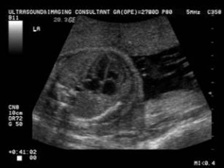
LEFT AND RIGHT
VENTRICLES THE INTERVENTRICULAR
SEPTUM |
- Ventricular Proportion. The
RV is slightly larger than the
- Ventricular Concordance (ventricles are connected to their respective atria).
Heart – cross-sectional diameter (Table)– Jeanty et.al. 1984
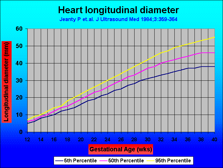
Heart – longitudinal diameter (Table)–
Jeanty et.al. 1984
Heart – cross-sectional diameter (Graph)– Jeanty et.al. 1984
Heart – longitudinal diameter (Graph)–
Jeanty et.al. 1984
- Left Ventricle.
- Closest ventricle to the spine.
- Identifiable structures include ventricular trabeculation, anterior and posterior papillary muscles, chordae tendinae and occasionally aberrant ventricular bands (false tendon) (1).
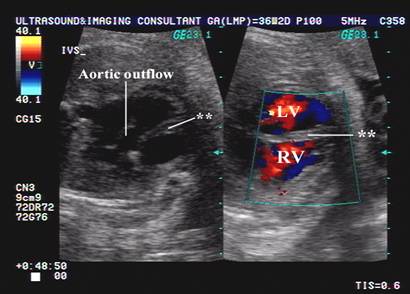
- Aorta arising from
- Normal
Size and configration of the left ventricle.
Left ventricle cross sectional diameter (diastole) (Table)– Wladimiroff et.al. 1982
Left ventricle cross sectional diameter (systole) (Table)- Wladimiroff et.al.
1982
Left ventricle cross sectional diameter (diastole) (Graph)– Wladimiroff et.al. 1982
Left ventricle cross sectional diameter (systole) (Graph)- Wladimiroff et.al. 1982
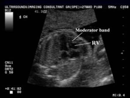
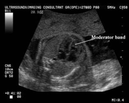
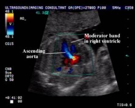
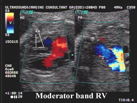
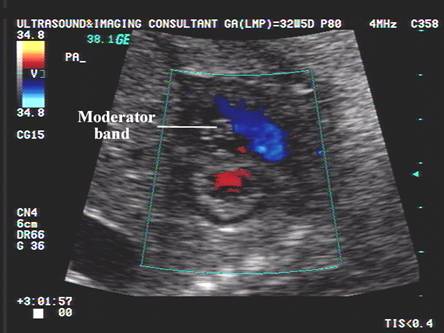
- Right Ventricle.
- Contacts the anterior chest wall.
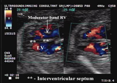
- Has a muscular (moderator) band near its apex (2).
-
- Other identifiable structures include ventricular trabeculation septal papillary muscles, chordae tendinae, anterior and posterior papillary muscles.
- Normal
Size and configuration of the right ventricle.
|
|
Video clip of
moderator band of the right ventricle
|
|
|
|
|
|

Right ventricle cross sectional diameter (diastole) (Table)- Wladimiroff et.al.
1982
Right ventricle cross sectional diameter (systole) (Table)- Wladimiroff et.al.
1982
Right ventricle cross sectional diameter (diastole) (Graph)- Wladimiroff et.al.
1982
Right ventricle cross sectional diameter (systole) (Graph)- Wladimiroff et.al.
1982
|
|
|
|
|
|
|
|
|
|
|
|
|
Right
ventricle: ·
Situated anteriorly.
|
|
- Echogenic
Intracardiac Foci
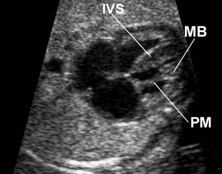
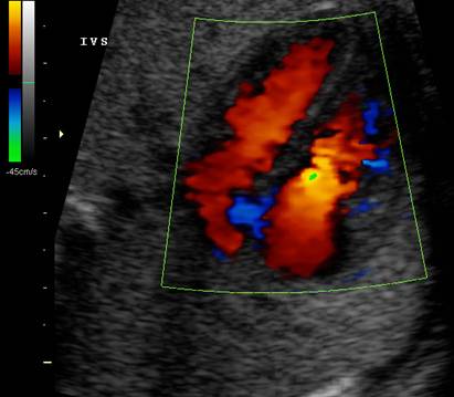
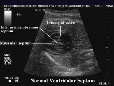
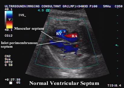
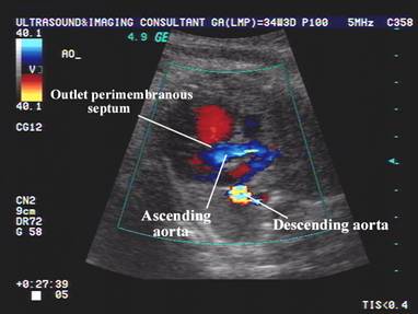
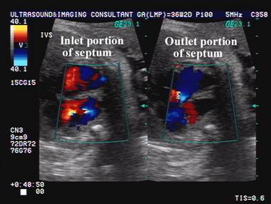
- Interventricular
Septum.
|
|
|
|
|
|
|
|
|
|
|
|
_files/Heart%20LV%20cross%20sectional%20diameter%20(diastole)_14251_image001.gif)
_files/Heart%20LV%20cross%20sectional%20diameter%20(systole)_19783_image001.gif)
_files/Heart%20RV%20cross%20sectional%20diameter%20(diastole)_31573_image001.gif)
_files/Heart%20RV%20cross%20sectional%20diameter%20(systole)_24540_image001.gif)
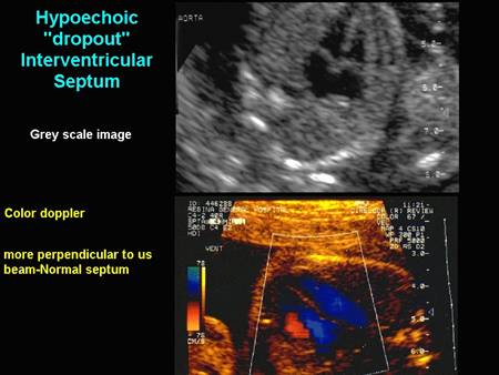
Interruption of the interventricular septum may be due to echo "dropout"- occurs if the septum is aligned parallel to the long axis of the ultrasound beam. Change transducer angle so that the septum is perpendicular to ultrasound beam.