Type
II Osteogenesis Imperfecta
|
|
|
|
|
Note
the exquisite visualization of the near field choroid plexus in the fetus
with absent cranial ossification.
|
|
3.
Femora >3SD below the expected mean femoral length for that gestational
age
(severe micromelia).
4.
Bones are short and broad.
|
|
Small thick, bowed femur
|
|
|
|
|
|
Radius + ulna (3D)
|
|
|
Tibia + fibula (3D)
|
|
Tibial fracture (2D and 3D)
|
|
|
|
Humerus
– micromelia + acute angulation due to fracture
|
|
|
|
|
|
|
|
14 wks
34 wks
|
|
9.
Pseudotalipes – due to the bowing and
fractures.
|
|
10.
Spine:
a.
Platyspondyly.
b.
Normal ossification.
|
|
|
|
|
|
|
|
|
|
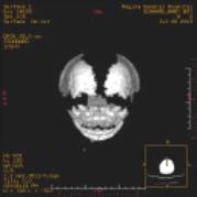
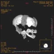
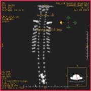
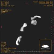
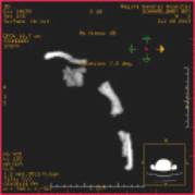
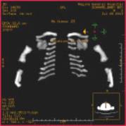
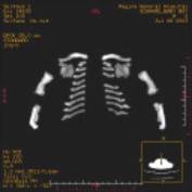
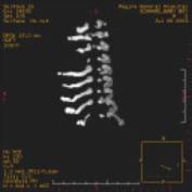
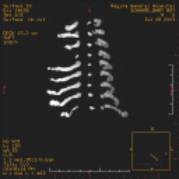
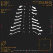
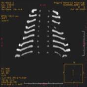
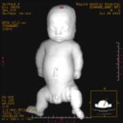
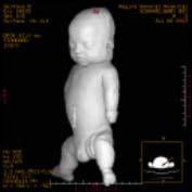
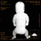
3D post
mortem CT scan
|
|||
|
|
|
|
|
Plain Films
|
|
|
|
|
|
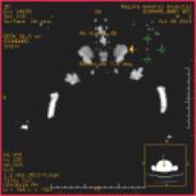
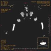
Coronal CT images
|
|
|
|
|
Cranium
|
|
|
|
|
Pelvic Girdle
|
|
|
|
|
Spine (left)
Upper limbs (right) |
|
|
|
|
Thoracic Cavity
|
|
|
|
|
|
|
|
|
|
|
|
|
DIFFERENTIAL DIAGNOSIS OF LETHAL FORMS |
DIFFERENTIAL DIAGNOSIS OF NON - LETHAL FORMS |


