The Normal
Pelvic Ligaments
Not routinely seen in the absence of
fluid in the pelvis.
|
- Broad Ligaments
- Double fold of
peritoneum that arises from the mullerian
ducts.
- Loosely
holds the uterus, fallopian tubes and round ligament in their normal positions,
suspending them from the lateral pelvic walls
|
- Round Ligaments
of the Uterus
- Narrow flat
bands.
- 10-12cm long.
- Lie between the
layers of the broad ligament and are antero-inferior
to the fallopian tubes.
- Provide
some support by attaching the uterus to the anterior peritoneal wall,
and traveling through the inguinal canal to the labia majora.
|
|

|
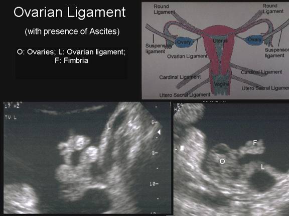
- Ovarian Ligament
- Rounded cord.
- Extends from the
superior angle of the uterus to the ovarian hilus.
- Seldom seen
during routine scanning.
|
- Transverse
Cervical Ligament (Mackenrodt or cardinal
ligament).
- Extends
laterally from the side of the cervix to the lateral fornices of the vagina.
- Together with
the uterosacral ligaments, it helps stabilize
the uterus from below.
- Seen
occasionally as a hypoechoic band that
appears to drape over the cervix.
|
|
|