|
ANATOMY OF THE SMALL
INTESTINES |
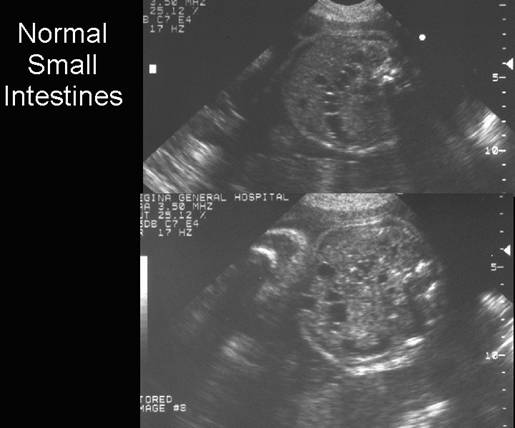
In early gestation (10-20 weeks), the small bowel lumen is difficult to demonstrate. The region of the small bowel appears as a hyperechoic area (relative to the fetal liver), in the abdomen and pelvis. This hyperechoic appearance around the small bowel persists throughout pregnancy. Between 12-16 weeks, this hyperechoic area occupies a large portion of the fetal pelvis and lower abdomen.
Criteria for normality include:
- No shadowing from the hyperechoic area.
- No associated cystic masses or other abnormalities.
- No ascites or polyhydramnios.
As the gestation progresses, the hyperechoic area becomes less prominent and is more centrally located in the fetal abdomen (1).
The hyperechoic appearance is thought to be due to:
- Reflections from walls of collapsed loops of small bowel.
- Mesenteric fat between loops of collapsed small bowel, as later in gestation, spaces between loops of fluid-filled small bowel also appear hyperechoic.
This hyperechoic area may be unusually prominent and should not be mistaken for a pathologic mass (2,3). Follow up scans in a few weeks will show a change in appearance or disappearance of this hyperechoic area (1).
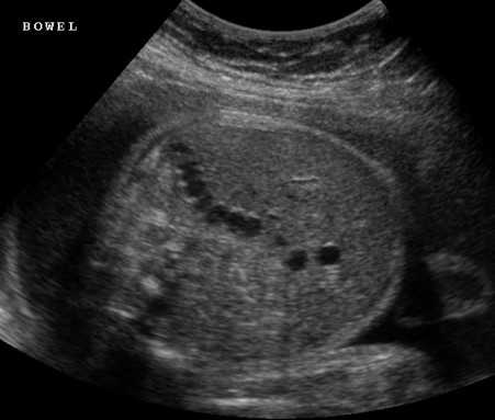
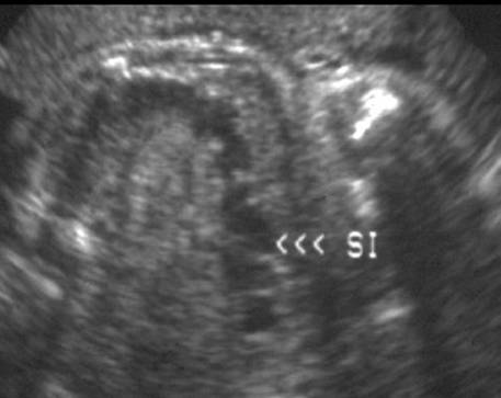
Between 13-20 weeks, hypoechoic rounded areas may develop within this hyperechoic region and probably represent small bowel lumen. Later in pregnancy, longer segments of fluid-filled small bowel loops can be demonstrated as rounded areas, however valvulae conniventes are only visualized in a small percentage of patients (1).
- Individual segments should
be:
£ 7-10 mm in diameter.
15 mm in length.
|
|
|
|
|
|
- Peristalsis (shifting of hypoechoic fluid) can be seen as early as 25 weeks.
|
GA (wks) |
Small Bowel Peristalsis |
|
10-15 |
0% |
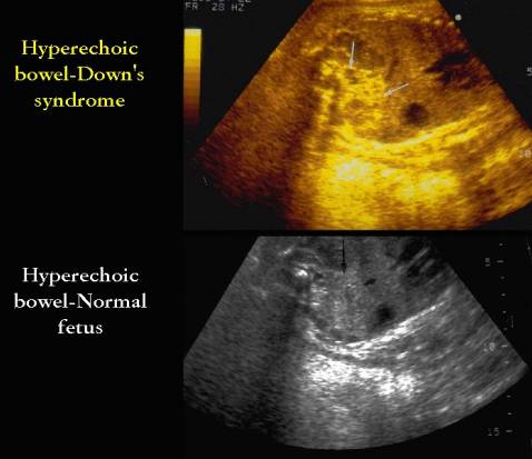
- Hyperechogenic bowel may be pathologic in
some cases.
|
|
REFERENCES |
- Parulekar SG. Sonography of normal fetal bowel. J Ultrasound Med 1991;10:211-220.
- Fakhry J, Reiser M, Shapiro LR et.al. Increased echogenicity in the lower fetal abdomen: a common normal variant in the second trimester. J Ultrasound Med 1986;5:489.
- Lince DM, Pretorius DH, Manco-Johnson ML. The clinical significance of increased echogenicity in the fetal abdomen. AJR 1985;145:683.