|
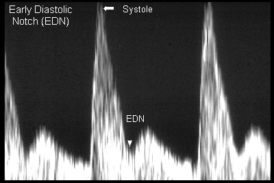
EARLY DIASTOLIC NOTCH
(EDN) OF THE UTEROPLACENTAL FLOW
VELOCITY WAVEFORM |
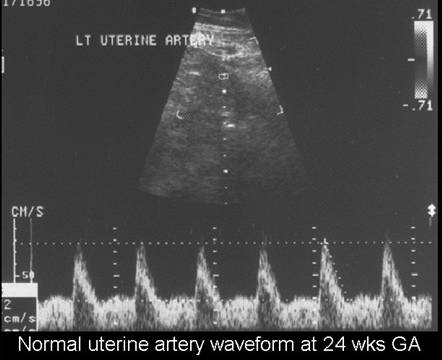
- Measurement. Uterine artery is visualized at its apparent point of crossing the external iliac artery on the pelvic sidewall. Doppler gate is placed medial to the external iliac artery to obtain a signal unaffected by this vessel. Signals obtained in mid trimester should be averaged out, as lateralization of placenta is difficult to identify on ultrasound. There is still controversy as to whether transabdominal or endovaginal scans are more reliable.
|
|
|
|
Video clip of
Uterine Artery Waveform
|
|
|
|
|
|

- Probably represents a reflective wave.
- Indicative of a high resistance circulation.
- Thaler
- EDN in uterine artery FVW in 50% of first trimester pregnancies.
- EDN disappeared by 26 weeks in normal pregnancies.
- Disappearance by end of second trimester probably relates to loss of elastic smooth muscle elements from spiral arteries (normal physiological change).
|
|
- EDN is present in at least one of the uterine artery FVW at the beginning of the majority of normal pregnancies.
- Bilateral notches are uncommon from 18 weeks onward.
- Grade 1 (mild) - lowest
part of notch < half of diastolic frequency shift.
Grade 2 (severe) - deepest part of notch < half of diastolic flow. - Abnormal
- Increase in one of the flow indices in early second trimester.
- Notching or increased indices in late pregnancy.
- Predispose to PET and IUGR.