|
THE INNER CELL MASS |
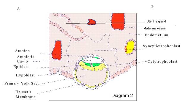
- Around the time of implantation, the inner cell mass differentiates into the bilaminar embryonic disc, consisting of an upper epiblast and lower hypoblast.( Diagram 2A)
|
|

- The amniotic cavity is found dorsally in reference to the embryonic disc. The primordial embryonic cavity is hollowed out within the pre-epithelial epiblast and is covered by cells derived from the inner cell mass (Diagram 2).
|
|
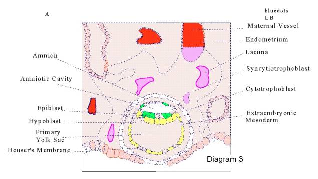
- The roof of the amnion then opens, exposing the primordial cavity to the overlying trophoblast. Around the 8th day post fertilization, amniotic epithelium migrates along the inside of the amniotic cavity, thereby forming the inner lining. The outer surface of the amniotic cavity becomes covered with extraembryonic mesoderm.( Diagram 3A)
|
|
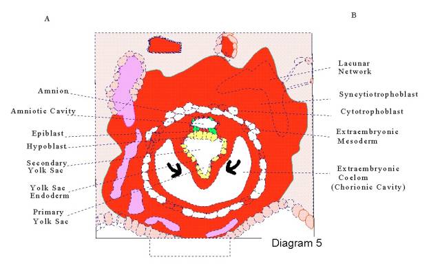
- The primary yolk sac (extracoelomic cavity) is found ventrally in reference to the embryonic disc, and is lined by an exocoelomic (Heuser's) membrane. This membrane is made up of flattened cells originating from the cytotrophoblast, and are replaced by the end of the second week as the hypoblast produces endodermal cells that migrate along the inside of the exocelomic membrane. The endodermal cells proliferate and form a secondary yolk sac within the new extraembryonic coelom.( Diagram 5A & 6A)
|
|
|
|
|
|

- The appearance of amniotic ectodermal epithelium and yolk sac endoderm is secondary to Gastrulation, occurring between the first and third weeks. It is this process that establishes the three germ layers.
- By the third week the amnion is a tough, thin, nonvascular membrane and is attached to the dorsal periphery of the now trilaminar disc.
- Around the fourth week, the
formation of the primordial nervous system (neurulation) and cephalocaudal
flexion of the embryo results in a tubular embryo with the amnion
attachment restricted to the umbilical area of the ventral wall.
( Diagram 7)
|
|
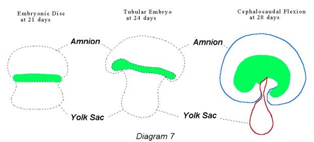
- The portion of the amnion nearest the umbilicus attaches to form an external covering over the newly developed cord. The initial wide communication between the embryo and the yolk sac becomes constricted until only a narrow, long, vitelline duct remains.( Diagram 7 & 8)
|
|

- The amniotic cavity enlarges rapidly as the amnion expands at the expense of the extraembryonic coelom. By the end of the third month, it has filled the chorionic sac and has loosely fused with the chorionic wall. Fusion occurs first with the decidua capsularis and then with the decidua parietalis.