THE
|
- The primary yolk sac develops due to growth of the extra-embryonic ectoderm from the ventral aspect of the embryonic disc. It cannot be visualized sonographically and soon degenerates and is replaced by the secondary yolk sac.
- First structure visible within the gestational sac and is of embryonic origin. The appearance of a yolk sac within the gestational sac excludes a blighted ovum pregnancy and ectopic pregnancy (rare cases of heterotopic pregnancies are however not excluded).
- Always seen when gestational sac = 13mm.
- Seen at 5 weeks MA when sac = 4mm in diameter.
- Confirms that the fluid collection within the uterus is a pregnancy as the double decidual reaction is not 100% specific for the presence of an intrauterine pregnancy.
|
ULTRASOUND |
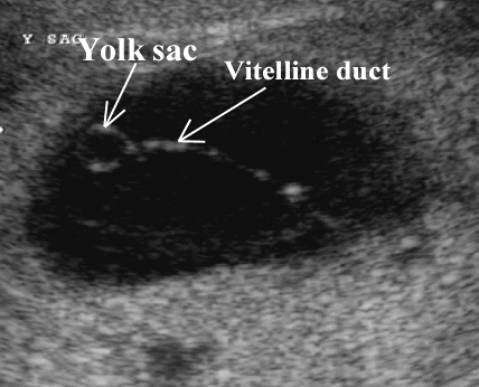
- Round or spherical with a bright echogenic rim and internal anechoic area.
|
|
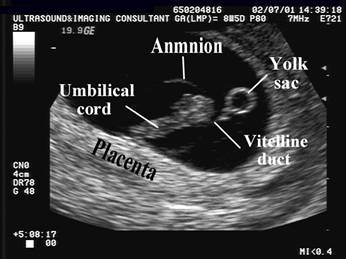
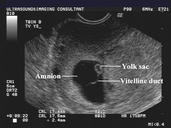
- It is localized outside the amnion within the chorionic space.
- 4-7 mm between 5 - 10 weeks GA (1).
|
Mean yolk sac
diameter during the first trimester |
|
|
Gestational age (weeks) |
Sonographic diameter (mm ±SD) |
|
5 |
3.01
± 0.75 |
|
6 |
2.99
± 0.73 |
|
7 |
3.99
± 0.86 |
|
8 |
4.72
± 0.64 |
|
9 |
5.22
± 0.63 |
|
10 |
5.89
± 0.56 |
|
11 |
5.35
± 0.87 |
|
12 |
4.34
± 0.62 |
- Growth = 0.1mm per millimeter growth of MSD when MSD <15mm. 0.03mm per millimeter growth of MSD thereafter.
- Situated in extraembryonic celom.
- Precedes visualization of the embryonic pole by 3-7days.
|
|
|

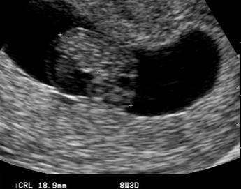
- Develops concurrently with the embryonic plate.
- Persists until about 14 weeks gestation (obscured by gestational sac and fetus).
- Wall thickness.
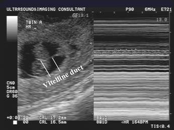
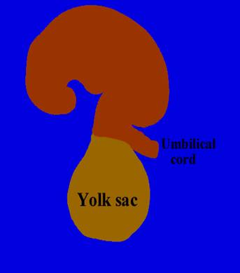
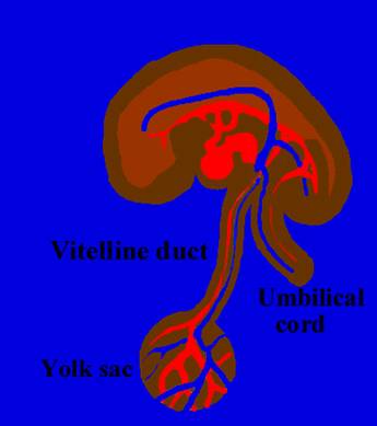
- Yolk sac remains connected
to the fetal midgut by
the vitelline duct.
|
|
|
|
|
|
|
|
|

|
|
|
|
|
|
|
|

- Function (2,3).
- Transfer of nutrients to the developing embryo at 3-4 weeks.
- Hematopoeisis occurs in the wall in the 5th week prior to this function being taken over by the fetal liver in week 8.
- Dorsal part of the yolk sac is incorporated in the embryo as the primitive gut in week 6.
- Usually disappears by the end of the 12th week. A recent study suggests that instead of being compressed, it degenerates first and disappears as a result of involution rather than mechanical pressure (3). Doppler studies (4) demonstrate a decrease in yolk sac vascularity after 9 weeks of gestation.
- The presence of a normal yolk sac has been associated with a 62% incidence of a normal pregnancy (5).
REFERENCES
|
- Manton M, Pederson JF. Ultrasound visualization of the human yolk sac. J Clin Ultrasound 1979;7:459.
- Jolly WP. Development, morphology and function of the yolk sac placenta of laboratory rodents. Tetralogy 1990;41:361-366.
- Janiaux E, Jurkovic D, Henriet Y. Development of the secondary human yolk sac: correlation of sonographic and anatomical features. Human Reprod 1991;6:1160-1165.
- Kurjak A, Kupesic S, Kostovic L. Vascularization of the yolk sac and vitelline duct in normal pregnancies studied by transvaginal color and pulsed doppler. J Perinat Med 1994;22:433-440.
- Nyberg DA, Laing FC, Filly RA. Threatened abortion: Sonographic distinction of normal and abnormal gestational sacs. Radiology 1986;158:397.