Adrenal length – left and right side
(Table) – Bronshtein et.al. 1993
Adrenal length – right side (Graph)–
Bronshtein et.al. 1993
Link to Embryology
- Large echolucent
cortex surrounding an echogenic medulla. The
normal appearance of the adult adrenal gland is the reverse.
- May be confused with the
kidneys at 9 weeks GA as they are of similar size and in the same area of
the abdomen.
- They can be imaged at the
end of the first trimester but are only reliably visualized after 20 weeks
gestational age (1,2).
- Their size increases
linearly from 12-17 weeks of gestation (3).
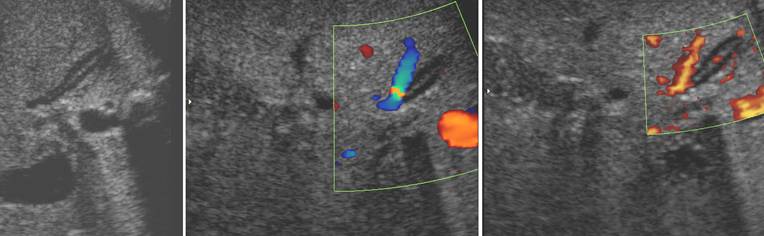
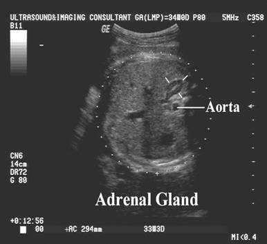
- They lie superior and
medial to the corresponding kidney, with the medial border adjacent to the
aorta (on the left) and inferior vena cava (on the right).
- Width
of adrenal limb <4mm
- Smooth
surface
- Echogenic central stripe with surrounding hypoechoic rim (6-8).
- Echogenic central stripe – represents the
congested sinusoids of the inner part of the fetal adrenal cortex, the
central vein and small medulla.
- Peripheral
hypoechoic area represents the less congested
adrenal coetex and probably the definitive
cortex.
- Shape.
- Oval or pyramidal
shaped in a longitudinal plane.
- In a transverse plane
they have been compared to radially oriented
rice grains (lentiform or discoid).
- The internal echogenic area may be quite bright in two thirds of
fetuses and may get even brighter in the last 5 weeks of pregnancy (1,2).
- Length increases with age,
and it maintains a constant relationship to kidney length (2).
- 1.4 to 2.2 cm or
48-66% of the renal length (4).
- The adrenal gland
constitutes 0.2% of total body weight at birth (this is 20 times it's relative size in the adult) (4).
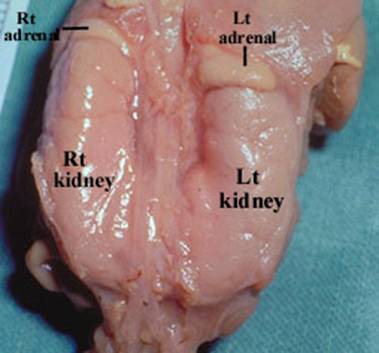
- Renal agenesis.
- The adrenals assume
a reniform shape making the early
diagnosis of renal agenesis difficult.
- Adrenals may
completely fill the renal fossa.
- The adrenal glands
may appear more globular and simulate small but present kidneys. Autopsy
studies have shown this phenomenon to be caused by a change in the
normal-sized adrenal’s shape, rather than adrenal hypertrophy (9).
Looking at orthogonal images may avoid this problem (10).
- Hoffman et al (11)
reported a flat or "lying down" adrenal in 48% of 23 fetuses
and 6 neonates retrospectively studied by US because of apparent renal
agenesis or ectopia and suggested that the
normal shape of the adrenal gland which usually "caps" the
kidneys is affected by presence of an ipsilateral
kidney.
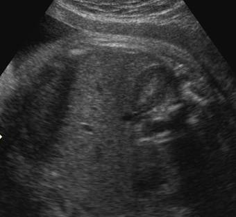
Discoid adrenal gland in unilateral renal agenesis
|
|

|
|
|
- Jeanty
P, Chervenak F, Grannum
P et.al. Normal ultrasound size and
characteristics of the fetal adrenal glands. Prenat
Diagn 1984;4:21.
- Lewis E, Kurtz A, Dubbins P et.al. Real time ultrasonographic evaluation of normal fetal adrenal
glands. J Ultrasound Med 1982;1:265.
- Bronshtein
M, Tzidony D, Dimant M
et.al. Transvaginal
ultrasonic measurements of the fetal adrenal glands at
- weeks
of pregnancy. Am J Obstet Gynecol
1993;169:1205-1210.
- Lee W, Comstock CH, Jurcak-Zaleski S. Prenatal diagnosis of adrenal
hemorrhage by ultrasonography. J Ultrasound Med
1992;11:369-371.
- Hauffa
BP, Menzel D, Stolecke
H. Age related changes in adrenal size during the first year of life in
normal newborns, infants and patients with congenital adrenal hyperplasia
due to 21-hydroxylase deficiency: comparison of ultrasound and hormonal
parameters. Eur J Pediatr
1988;148:43-49.
- Scott EM, Thomas A, McGarrigle HHG, Lachelin
GCL. Serial adrenal ultrasonography in normal
neonates. J Ultrasound Med 1990;9:279-298.
- Kangarloo
H, Diament MJ, Gold RH et.al.
Sonography of adrenal glands in neonates and
children: changes in appearance with age. J Clin
Ultrasound 1986;14:43-47.
- P. Dubbins,
A. Kurt, R. Wapner et al., Renal agenesis:
spectrum of in utero findings. J Clin Ultrasound 9 (1981), pp.
189–193.
- S. Droste,
J. Fitzsimmons, J. Pascoe-Mason et al., Size of the fetal adrenal in
bilateral renal agenesis. Obstet Gynecol 76
(1990), pp. 206–209.
- C.K. Hoffman, R.A. Filly
and P.W. Callen, The "lying down"
adrenal sign: A sonographic indicator of renal
agenesis or ectopia in fetuses and neonates. J
Ultrasound Med 11
(1992), pp. 533–536.
