|
BRANCHIAL CLEFT CYSTS
AND REMNANTS |
Branchial cleft cysts are congenital epithelial
cysts, which arise on the lateral neck from a failure of obliteration of the
second branchial cleft in embryonic development.
EMBRYOLOGY |
Brachial remnants (cysts, fistula, skin tags, chondrocuteneous
vestige) are developmental anomalies of this system. Cervical chondrocutaneous
branchial remnants are usually embedded in the anterior border of the
sternocleidomastoid muscle and appear as skin tags.
At the fourth week of embryonic life, the development of 4 branchial (or pharyngeal) clefts results in 5 ridges known as the branchial (or pharyngeal) arches, which contribute to the formation of various structures of the head, neck, and thorax. The second arch grows caudally and ultimately covers the third and fourth arches. The buried clefts become ectoderm-lined cavities, which normally involute around week 7 of development. If a portion of the cleft fails to involute completely, the entrapped remnant forms an
These four arches are separated by four paired clefts and pouches.
1. The first
pharyngeal pouch gives rise to the eustachian tube, middle ear and mastoid
cells.
2. The
second pouch forms the supratonsillar fossa.
3. The third pouch develops into the thymus gland and upper parathyroid glands.
4. The fourth pouch gives rise to the lower parathyroid gland and part of the thyroid gland.
LOCATION |
There are four different types of cysts classified according to their
location. Superficial cysts are found anterior to the sternocleidomastoid
muscle. Cysts found anterior to the carotid artery and jugular vein are most
frequently encountered. Another group of cysts are found extending to the
pharynx and the final group are those cysts that extend between the vessels and
the lateral wall of the pharynx.
The first branchial cleft cysts are generally located in
the lower pole of the parotid gland and are often first noticed in middle age.
A branchial cleft cyst is the most common branchial abnormality
with 80–90% arising from the second branchial cleft. Branchial cleft
cysts can occur anywhere from the mandibular angle (tonsillar fossa) to the
supraclavicular area
Anomalies arising from the third and fourth branchial cleft are uncommon but when present they present as fistulas of the pyriform sinus and abscesses.
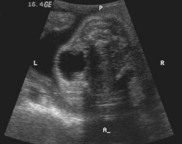
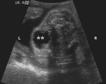
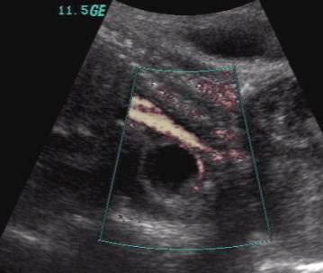
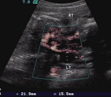
ULTRASOUND |
Uncomplicated cysts present as a recurrent, painless, fluctuant
and non-tender neck mass often seen in later childhood and adulthood. Prentation
in the fetus, although not common has been reported.
·
Well-defined
anaechoic, thin wall cystic mass that displaces the carotid sheath medially or
posterolaterally and the sternocleidomastoid muscle posteriorly or
posterolaterally.
·
May uncommonly
appear solid if the content is mucoid or contains cholesterol.
·
When
infected, the cyst usually acquires a thick wall with echogenic debris within,
in which case the cyst may then be surrounded by lymph nodes.
- Approximately 2-3% of cases are bilateral
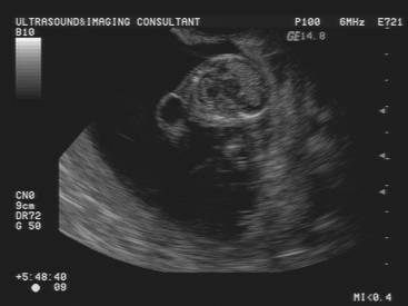
Case 1 |
|
|
|
|
|
|
|
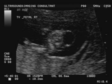
Case 2 |
|
|
|
|
REFERENCES |
1.
Suchet I Ultrasonography of the fetal neck in the second and third trimesters.
Part 3. Anomalies of the anterior and anterolateral nuchal region Can Assoc Radiol
J 1995; 46:426-433