|
CAPUT SUCCEDANEUM |
In prolonged labor, the dependent area of the fetal scalp
in immediate contact with the cervix may become edematous. A swelling, known as
the caput succedaneum develops (1). The caput may be formed with the fetal head
lower in the birth canal, commonly only after the fetal head encounters a rigid
vaginal vault (2).
ULTRASOUND |
- Prenatal detection has been described (1,3).
- Soft tissue mass overlying the cranium.
- Overlapping or "moulding" of the sutural bones.
- Color doppler differentiates it from a vascular hemangioma.
|
|
|
|
|
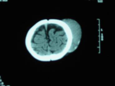
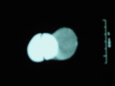
CT scan on Day 1
Notice
the large amount of soft tissue swelling overlying the left parietal bone. |

DIFFERENTIAL DIAGNOSIS |
- Encephalocele (4).
- Cephalhematoma - subperiosteal hematoma and does not cross the suture line.
- Caput succedaneum - focal swelling of the scalp from edematous fluid overlying the scalp and can therefore does cross suture lines.
- Hemangioma (vascular lesion).
PROGNOSIS |
- Resolves spontaneously.
REFERENCES |
- Sherer DM, Allen TA, Ghezzi F et.al. Enhanced transvaginal sonographic depiction of caput succedaneum prior to labor. J Ultrasound Med 1994;13:1005-1008.
- Cunningham FG, McDonald PC,
- Schiwmer SR, Lebovic J. In utero sonographic demonstration of a caput succedaneum. J Ultrasound Med 1986;5:711.
- Sanders RC. Obstetrics:
Fetal head and neck. In: Atlas of Ultrasonographic
Artifacts and Variants. 2nd ed.