|
THE YOLK SAC |
NORMAL YOLK SAC |
ABNORMAL YOLK SAC |
- Large yolk sac >6mm.
- Small yolk sac £2mm (1).
- Irregular shape.
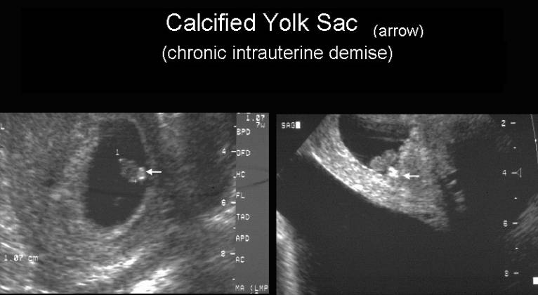
- Calcified yolk sac - due to longstanding embryonic demise.
- Floating yolk sac.
- Solid echodense yolk sac (associated with fetal death or an anomalous fetus) (1).
- Non visualization is always abnormal.
- Thin yolk sac - predictive value of 53.8% for an abnormal outcome.(2)
- Thick yolk sac - predictive value of 93.3% for an abnormal outcome.(2)
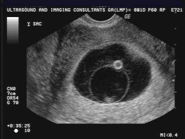
Dense, echogenic
yolk sac. Echogenic chorionic cavity
|
|||
|
|
|
||
|
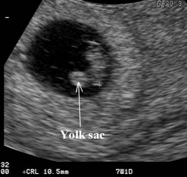
Small (1.6 mm)
yolk sac with an echogenic chorionic
cavity. |
|||
|
|
|
||
|
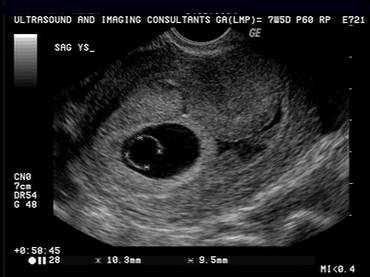
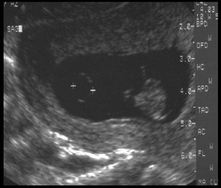
Large yolk sac (10.3 X
9.5 mm) at 7 weeks 5 days. Adjacent fetus seen, but amniotic cavity not yet visible. |
|||
|
|
|
||
|
|
|
||
|
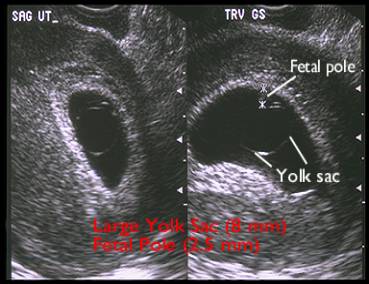
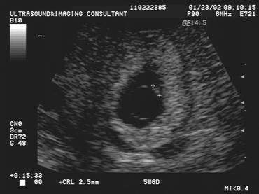
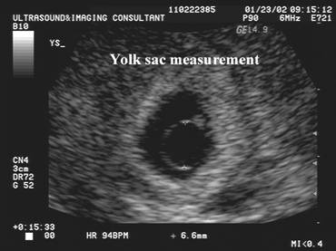
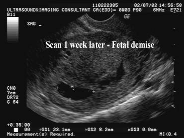
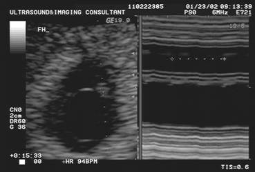
Large
yolk sac: -
yolk sac 6.6 mm -
fetal bradycardia 94 bpm - demise 1 week later |
|||
|
|
|
||
|
|
|
||
|
|
|
||
|
|
|||
|
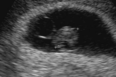
Floating Yolk Sac Thin
Yolk Sac |
|||
|
|||


REFERENCES |
- Green JJ, Hobbins JC. Abdominal ultrasound examination of the first trimester fetus. Am J Obstet Gynecol 1988,159:165.
- Levi CS,
- Ferrazzi E, Brambati B, Lanzani A. The yolk sac in early pregnancy failure. Am J Obstet Gynecol 1988;158:137-141.
- Recce EA, Scioscia AL, Ointer E. Prognostic significance of the human yolk sac assessed by ultrasonography. Am J Obstet Gynecol 1988;159:191-196.