|
COLOR DOPPLER IMAGING OF UTEROPLACENTAL CIRCULATION |
NORMAL PATTERN |
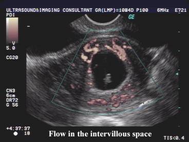
During the first 12-13 weeks of a normal gestation, the uteroplacental and
fetoplacental circulations are not in direct contact with each other (1,2).
There is no active blood flow through the decidual spiral arteries into the
intervillous space (3).
|
|
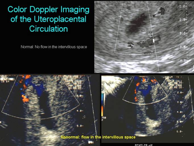
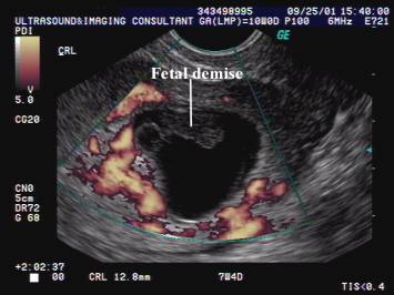
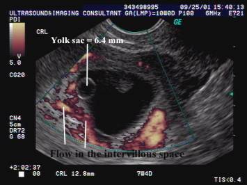
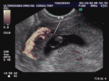
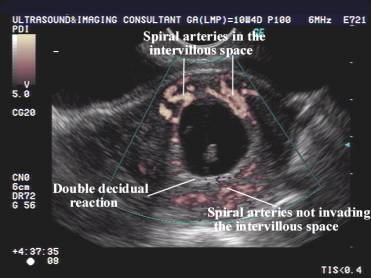
ABNORMAL PATTERN |
High resistance to blood flow within the decidual spiral arteries and the
presence of arterial blood in the intervillous space is associated with a high
risk of early miscarriage (4). This is thought to be due to abnormally
high-pressure flow on the immature placenta resulting in disengagement of the
early villi, and subsequent miscarriage.
Endovascular trophoblast invasion
has been considered to serve the "purpose" of establishing, from the
earliest days of gestation, a maternal circulation providing the conceptus with
nutrients. This circulation was conceived originally as a sluggish capillary
derived blood pool, which evolves with trophoblast remodeling of the spiral
arteries into a high-volume, low-resistance circuit.
Hustin and Schaaps have challenged this
theory based on their failure to identify intervillous circulation both on
direct visualization of the intervillous space and in perfused hysterectomy
specimens. They demonstrated occlusion of the uteroplacental circulation by
trophoblastic plugs until the twelfth week of pregnancy. They speculated that
these trophoblastic plugs protect the young conceptus from the force of
maternal arterial blood flow until implantation is well-established. Since
then, it has been proposed that precocious initiation of maternal arterial
perfusion of the intervillous space may be responsible for early pregnancy
loss. The issue of whether the entire period of embryogenesis occurs without
any direct contact with the maternal circulation has not been resolved. A
cogent rebuttal has suggested that fixation artifact and intervillous flow
rates (below the current limits of Doppler resolution) may explain most of the
observations. The issue of whether most or all of embryogenesis occurs in the
absence of contact with the maternal circulation awaits final resolution.
REFERENCES |
- Hustin J Schaaps JP. Echocardiographic and anatomic studies of the maternotrophoblastic border during the first trimester of pregnancy. Am J Obstet Gynecol 1987,157:162-168.
- Janiaux E, Burton GJ, Muscoso GJ, Hustin J. Development of the early human placenta: a morphometric study. Placenta 1991,12:269-276.
- Jaffee R, Woods JR. Color doppler imaging and in vivo assessment of the anatomy and physiology of the early uteroplacental circulation. Fertil Steril 1993,60:293-297.
- Jaffee R, Dorgan A, Abramowicz JS. Color doppler imaging of the uteroplacental circulation in the first trimester. AJR 1995,164:1255-1258.