|
ULTRASOUND IN ECTOPIC
PREGNANCY |
|
6.
Mimickers of Ectopic Pregnancy. 7.
Non-visualization of the Ectopic
Pregnancy on First Ultrasound. |
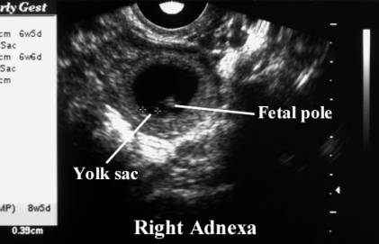
Definite ectopic pregnancy - an extra-uterine mass containing a definite fetal pole +/- a yolk sac +/- cardiac pulsations.
Suggestive signs of an ectopic pregnancy include (in order of highest likelihood):
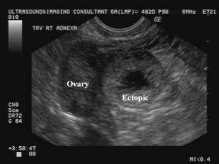
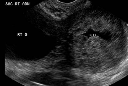
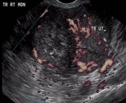
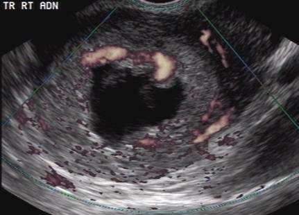
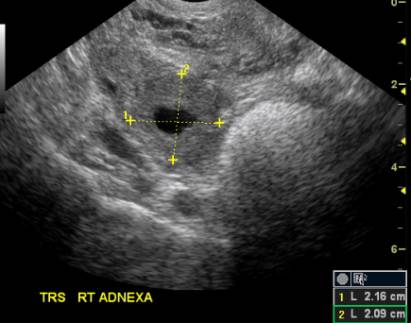
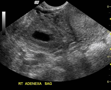
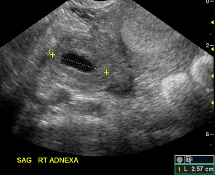
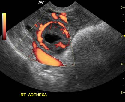
1. an empty uterus + a thick-walled adnexal mass separate from the ovary with an anechoic center that does not contain a yolk sac or fetal pole (> 95% likelihood).
2. an empty uterus + complex adnexal mass with solid and cystic components (> 70% likelihood) - especially if the adnexal mass is tender to probe palpation. an empty uterus + complex adnexal mass with solid and cystic components (> 70% likelihood) - especially if the adnexal mass is tender to probe palpation.
3. an empty uterus + moderate-large amount fluid in the cul de sac (> 50% likelihood).
(* a thin-walled cyst arising from the ovary is usually a simple corpus luteum cyst, but a hemorrhagic corpus luteum cyst may appear complex and may be ultrasonically distinguishable from a tubal ectopic pregnancy - which is a thick-walled cytic structure distinctly separate from the ovary - only by it's closer proximity to the ovary).
Indeterminate ultrasound is defined as an ultrasound showing no IUP and no signs suggestive of an ectopic pregnancy.
A patient with an indeterminate ultrasound may still have an ectopic pregnancy, with the degree of risk depending on the ultrasound findings:-
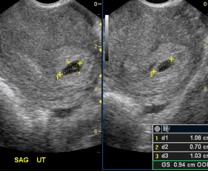
1. < 25% of patients with an empty uterus (no gestational sac) and no other findings will have an ectopic pregnancy.
2. echogenic material within the uterine cavity may be due to an anembryonic pregnancy or due to the presence of blood secondary to an incomplete abortion, but ~ 5% of cases are secondary to an ectopic pregnancy.
3. a patient with a normal-appearing gestational sac of 5 - 10mm in size + no other findings still has a small (< 5%) risk of an ectopic pregnancy
(* a patient with a serum HCG < 1,500 mIU/ml should still have an ultrasound performed at the time of initial presentation, because ~ 40% of ectopics with serum HCG levels < 1,000 mIU/ml can be identified by ultrasonography at the time of initial presentation)
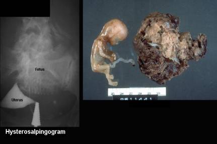
Location:
95% tubal
5% other
|
Ampulla
(75-80%) |
||
|
|
|
|
|
Isthmus
(10-15%) |
||
|
|
|
|
|
Fimbria (5%) |
||
|
|
|
|
|
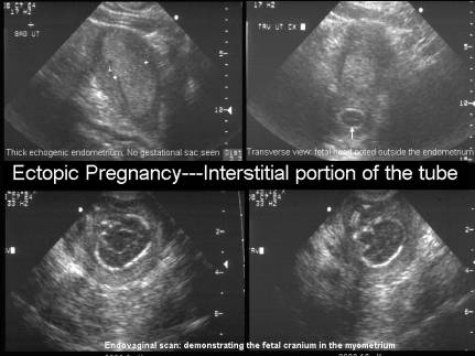
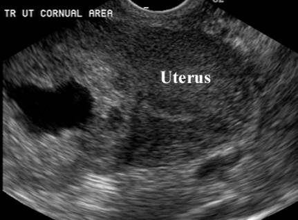
Cornual region
of uterine fundus (<5%) |
||
|
|
|
|
|
|
|
|
|
|
|
|
|
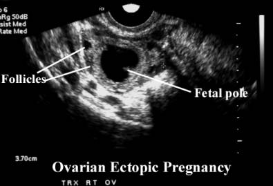
Ovary (<1%) |
||
|
|
|
|
|
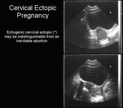
Cervix
(0.1%) |
||
|
|
||
|
Abdominal
pregnancy --intraperitoneal surface or between the leaves of the broad
ligament (0.03%) |
||
|
|
|
|
|
Heterotopic
pregnancies are rare – note the presence of a viable intrauterine and adnexal
pregnancy |
||
|
|
|
|
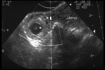
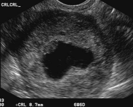
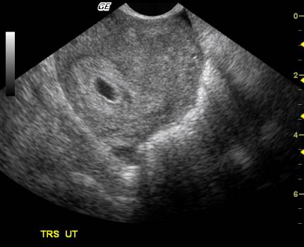
Small viable true intrauterine gestational sac |
||
|
|
|
|
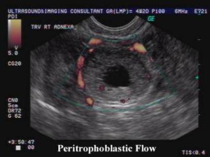
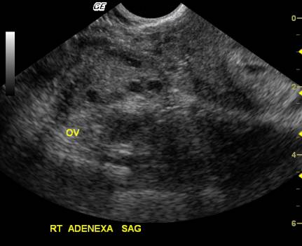
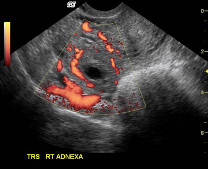
Adnexal ring separate from the ovary. Large amount of flow – “ring of fire” |
||
|
|
|
|
|
|
|
|